
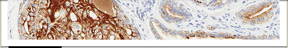
Preparation and analysis of virtual histological slides
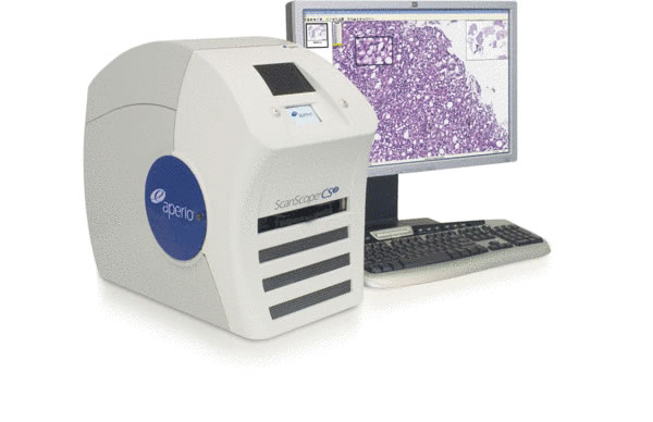 Virtual slides allow highly reliable storage, network sharing, and analysis and annotation of histological material. The Stem Cell Pathology Unit is equipped with two Leica/Aperio ScanScope systems. ScanScope CS2 capture device has a five-slide capacity and 20x and 40x magnification capabilities of bright field microscopy. ScanScope FL capture device allows preparation of whole slide images of slides stained with multiple fluorochromes. With the monochrome camera and quad multi-band pass filter set provided with the system,
 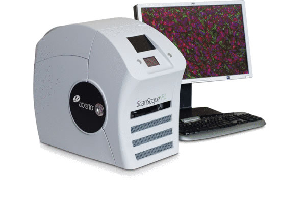 up to four color channels can be utilized.
Additional flexibility is provided by the six-position filter cube turret and excitation filter wheel. Advanced illumination, autoexposure, and autofocus capabilities eliminate the need for trial-and-error image capture, greatly minimizing the occurrence of photobleaching. These high-resolution multi-channel digital images can be archived permanently, eliminating concerns about fluorochrome fading on the glass slides.
Quantitative analysis of bright field and fluorescent images is performed on dedicated computers equipped with several software packages, such as Area Quant BF (Aperio area quantification software brightfield application), Whole Cell Quant (Aperio area quantification software brightfield application) and ToolboxFL (Includes Aperio Area Quantification FL, Indica Labs Cytonuclear FL, Indica Labs Object Colocalization FL, and Indica Labs Microvessel FL image analysis algorithms).